
AN INEXPENSIVE SOLUTION TO VIBRATION IN PHOTOMACROGRAPHY
originally published
Journal of Biological Photography Volume 66 No 3 July 1998
| Richard Norman, BS Retired Technical Associate Rochester Institute of Technology Rochester, NY 14623 |
Michael R. Peres, MS, RBP, FBPA Chair, Biomedical Photographic Communications Rochester Institute of Technology Rochester, NY 14623 |
INTRODUCTION
As many biomedical and technical
photographers have experienced, the workplace is often plagued by vibration
from a myriad of sources. The biomedical photography laboratories at the Rochester
Institute of Technology are no exception. The effects of vibration in these
situations results in the making pictures that are soft. The pictures that are
effected by vibration will not demonstrate the type of crispness that might
be expected.
Vibration may or may not be observed in the camera. With a space that might be needed for the illuminator, and a long bellows draw, a camera assembly that is suspended high is in an unstable position for imaging. This configuration is especially vulnerable to vibration. By the nature of how a macro system is used, the illumination, subject, and image forming modules are not rigidly locked together as is with the case of a modern compound microscope. This makes the system susceptible to vibration. Out hypothesis to try to and isolate the camera apparatus from the building by floating a platform using low-pressure air bags.
PARTS OF THE SYSTEM
The platform was designed using a 30-inch x 40-inch sheet of three-quarter
plywood. Under each corner was a 3-inch high block allowing space for the air
bag to partially expand. The air bags were inflatable rubber balls about the
size of soccer balls, cheap, flexible and obviously the type intended for young
children. Four balls were partially deflated and placed under the platform,
near the corners. To float the platform and apparatus, the bags were inflated
until each corner of the table was lifted a 1/16 of an inch off the base and
corner supports.
TESTING THE SYSTEM
True to Murphy's Law, the building would not vibrate when we wanted it to. To
induce vibration in the system, a battery-operated foot massager was firmly
pressed to a table leg for several seconds, always at the same place, but before
the shutter was released. It is relevant to mention that a camera's cable release
can transmit vibrations to a camera system if not carefully used.
Pictures were made at 36x on the film when the platform was resting on the blocks. Vibration was visually evident and can be observed in the photos that were included the original journal article. The lower photo in that article shows Cycas microsporophyll sporangia at 36x on the film with the platform floating. There was no evident vibration observed in the viewfinder however the image is evidently more clear. It should be noted that the observable vibrations might often be too subtle for observation. As a consequence of the photomechanical reproduction used in journals, the full extent of the results might also be masked.
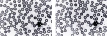
Following this experiment, it was decided to try a microscope with a magnifcation of 100x at the film plane. Contrary to expectation, no vibration was observable when the platform was resting on the blocks with the 10x objective. It was discussed that when photographing blood cells, vibration was often present. So when using a 100x oil immersion objective and a blood sample slide was selected, vibration was observed
The left picture shows loss of detail in the stained leukocyte, with the platform resting on the blocks with vibration, while the right picture shows 1000x on the film delineates crisper nuclear detail in the leukocyte, with the platform floating also with vibration.
CONCLUSION
The system described here produced a marked improvement in the results for a
macro application which was our original intentions.The
detail in the photomicrograph of the leukocyte showed improvement, but could
it be better yet? Was there still residual vibration? Probably yes. At these
high magnifications, the critical worker had better stick with a good commercial
vibration isolation table but the results shown here within demonstrate that
simple inexpensive solutions still have value.
ABOUT Richard N. Norman
Richard Norman retired from RIT in 1992. He is an active
member of Intermicro and is routinely working on solutions to problems found
in this area of photography. Many of his inventions can be found in the teaching
labs of the Biomedical Photographic Communications department.